The presentation documents treatment of a male patient with severe maxillary dental deterioration, esthetic compromise, and skeletal deficiencies requiring advanced full-arch implant rehabilitation. Initial facial photographs and clinical intraoral views demonstrated failing anterior restorations, severe dental deterioration, and compromised smile esthetics.
Preoperative skeletal CT scan views showed extensive maxillary defects and compromised alveolar support requiring advanced implant anchorage strategies. The pre-treatment intraoral views demonstrated multiple failing anterior teeth with inadequate esthetic and structural support.
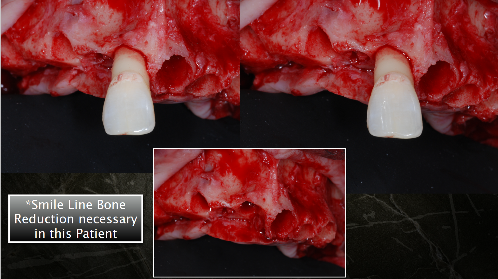
The surgical phase involved smile line bone reduction to establish restorative space and improve prosthetic positioning. Sequential intraoperative photographs showed:
- Surgical flap reflection
- Alveolar bone reduction
- Remaining socket depth measurements (~8 mm remaining)
- Preparation of implant osteotomy sites
Advanced implant reconstruction techniques included:
- Trans-sinus implant placement
- Bicortical stabilization using the lateral pyriform nasal plate
- Placement of maxillary All-on-6 implants
- Additional support with a single zygomatic implant
Subsequent surgical steps included:
- Bone grafting procedures
- Closure of osseous defects (“bone hole” closure)
- Continuous sling suture closure techniques
Postoperative 10-day CT scan and facial photographs demonstrated successful implant positioning, improved facial support, and healing progression. Final restorative treatment involved fabrication of a definitive milled monolithic zirconia full-arch prosthesis. Final “before and after” facial comparisons demonstrated substantial improvement in smile esthetics, facial appearance, and functional rehabilitation.