The presentation documents rehabilitation of the posterior left maxilla in a patient with insufficient posterior maxillary bone associated with sinus pneumatization. Pretreatment clinical photographs and CT panoramic imaging demonstrated compromised posterior maxillary dentition and reduced available bone volume in the sinus region.
Treatment began with atraumatic tooth removal to preserve residual alveolar bone and soft tissue architecture. A TempStent II surgical guide was used to confirm restorative positioning and implant angulation before osteotomy preparation.
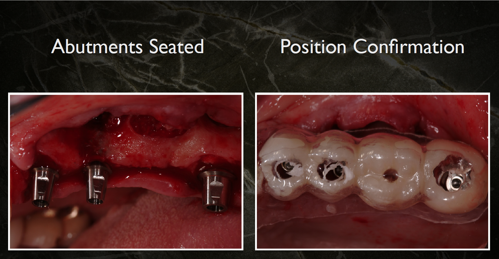
During sinus augmentation, separate pneumatized sinus pouches were identified and managed surgically to create adequate space for grafting and implant placement. Following implant insertion, abutments were seated and position confirmation was completed clinically using the restorative guide.
The restorative phase involved:
- Placement of provisional copings
- Correction and contouring of provisional restorations
- Immediate implant-supported provisionalization
- Immediate restoration of posterior occlusion and esthetics
Postoperative CT panoramic and serial CT images demonstrated successful implant positioning and sinus augmentation. Final five-year follow-up photographs and periapical radiographs documented:
- Stable peri-implant tissues
- Functional posterior implant restorations
- Maintenance of osseous support
- Long-term implant success and integration